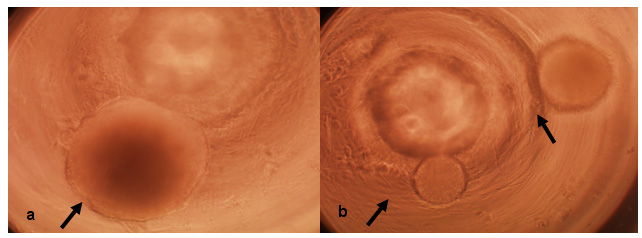
| Figure
2. Single
EB in 96-well format polycarbonate PCR plate. Results displayed
in a and a are phase contrast microscopy using the same magnification.
(a)
Single EB in a well. A single EB is present in the microwell and
the single EB is of typical size.
(b)
EB with dissimilar size and number. Two EB are present in the microwell
and the two EB are of dissimilar sizes. Additionally, sizes of the two
EB are dissimilar from the single EB displayed in a arrows point
to the EB. |