| Figure
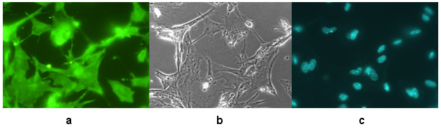
3. Characterization of ES cells differentiated into neurons on day 12
of differentiation.
(a)
Cells stained with neuronal marker. Cells show indirect immunofluorescence
of Oregon Green-conjugated antibody against the neuronal cell marker,
cytoskeletal protein MAP2 [a + b]. Cells that fluoresce are MAP2 positive
and display the neuronal cell phenotype.
(b)
Cell showing neuronal morphology. The same field of cells shown in (a)
are displayed using bright field microscopy. Characteristic neuronal
morphology is evident.
(c) Cells stained with DAPI showing the nucleus. The same field of cells shown in (a) and (b) is displayed as DAPI-staining
of the nucleus. |