| Figure
1.
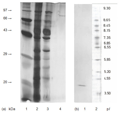
Analysis of purified β-glucosidase from Melanocarpus sp.
MTCC 3922.
(a)
SDS-PAGE of purified Lane 1, standard protein markers in the order of
increasing molecular mass: Soyabean trypsin inhibitor (20 kDa);
Carbonic anhydrase (29 kDa); Ovalbumin (43 kDa); Bovine serum albumin
(66 kDa); Phosphorylase b (97.4 kDa); Lane 2, crude protein; Lane 3,
protein from pool 1 of DEAE-Sepharose column; Lane 4, protein of purified
β-glucosidase after PBE-94 column.
(b)
Iso-electric focusing of purified β-glucosidase, Lane 1, protein of purified β-glucosidase after PBE-94 column and Lane 2 standard
pI markers. |