
|
|
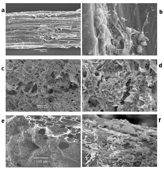
Figure 1. Scanning electronic micrographs of support materials: (a) sisal fibre waste (mag. x 500), (c) pumice stone (mag. x 35) and (e) porous glass beads surface and cross section (mag. x 150) before microbial colonisation, and after colonisation with anaerobic microbial biofilms: (b) sisal fibre waste (mag. x 6000), (d) pumice stone (mag. x 3000) and (f) porous glass beads (mag. x 2300). |
|
|