
|
|
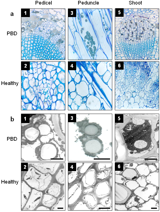
Figure 3. (a) Optical and (b) Electron microscopy analysis of pedicels, peduncles and shoots from plants exhibiting PBD. Ovoid and spherical bodies can be seen in the cytoplasm of phloem vascular system cells. Samples from healthy plants are included as negative controls. Viral particles were also observed (data not shown). a1: 20X, a2-4: 40X, a5-6: 10X. Bars: 1 µm (b1, b3, b5), 5 µm (b2, b4, b6). |
|
|