|
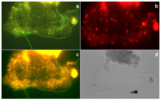
Figure 5. FISH staining of the sludge sample
analyzed by confocal laser microscopy of FISH cells (a, b and c) and
light microscopy (d).
(a) Methanosaeta concilii;
(b) Bacteria; (c) Simultaneously hybridized with rhodamine-labeled
bacterial-domain probe (EUB338) (red) and FITC-labeled methanogens probe
(MSMX860) (green) showing the consortium between methanogens and bacteria; (d)
Light microscopy. |