
|
|
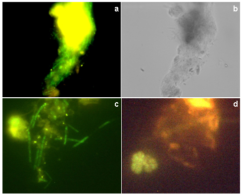
Figure 6. FISH staining of the sludge samples analyzed by confocal laser microscopy of fluorescent in situ -hybridized cells (a, c and d) and light microscopy (b). A fluorescent in situ -hybridized floc of Methanosaeta concilii (a); light micrograph of a floc of Methanosaeta concilii (b); filaments of Methanosaeta concilii (c); simultaneously hybridized with rhodamine-labeled bacterial-domain probe (EUB338) (red) and FITC-labeled methanogens probe (MSMX860) (green) showing Methanosarcina (d). |
|
|