
|
|
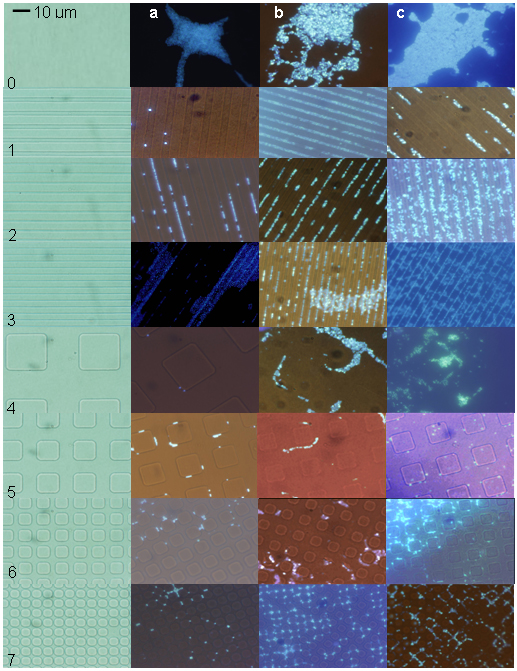
Figure 2. Features imprinted into silicone elastomer and growth of S. aureus (a), S. epidermidis (b), and P. aeruginosa (c) on imprinted silicone elastomer after 20 hrs. Patterns (bright field images) are identified in the left-most column and growth of individual organisms is shown in adjacent columns (combination epifluorescence and bright field images). Controls (0) were smooth surfaces and patterns consisted of bars (1, 2, and 3), squares (4, 5, and 6) and circles (7). Dimensions of features and spaces between features are given in Table 1. |
|
|