
|
|
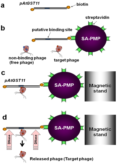
Figure 1. Schematic diagram of screening. (a) Both sides of the DNA probe (600 bp upstream region of the AtGST11 gene) were 3`end-labeled with biotin by a terminal deoxynucleotidyl transferase. (b) The DNA probe was captured with SA-PMP in the reaction solution and recombinant phages were added to the solution to make DNA/phage complex. (c) The complex was trapped to a magnetic stand and rinse with a washing solution to remove non-binding phages. (d) DNaseI treatment was performed to release the phages from the binding site. These steps (a to d) were repeated three times to increase the number of candidates in the population. |
|
|