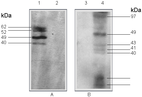
Figure 3. Western-blot
detection of hrIDS expressed in E. coli using an anti-hIDS monoclonal
antibody and ECL detection.
A: (Western-blot
from 8% w/v SDS-PAGE).
B: (Western-blot
from 12% w/v SDS-PAGE). 1A:
JM109/pUC13-hrIDS cell lysate, 2A: cell lysate, 3B: culture supernatant,
4B: culture supernatant and JM109/pUC13.
|