
|
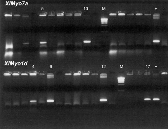
| Figure 3. SP screens for two myosin isoforms. SPs from step 3 of Figure 1 were screened in two separate sets of PCR reactions to identify SPs containing 7a cDNA clones (top lanes) or 1d (bottom lanes). Using 1 µl as a template per PCR, 17 of the 20 SPs were screened. Lanes designated M contain the same markers as in Figure 2 loaded at a lower concentration. The smallest visible band corresponds to the 506, 517 bp doublet. The 7a screen identified two positive SPs, numbers 5 and 10, and the 1d screen identified four SPs, numbers 4, 6, 12, and 17. Lanes designated (+) contain PCR reaction product in which the cloned DNA fragments from which the primer pairs were derived were used as template. Lanes () contain products of PCR reactions with no added DNA template. |
|
|