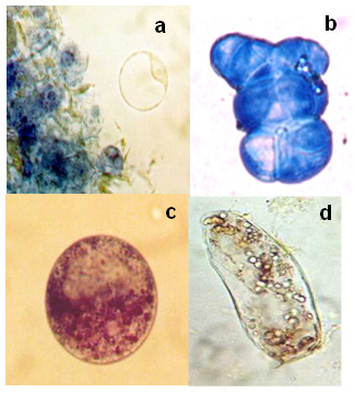
| Figure 1.
Viable
and non-viable cells and protoplasts stained with Evans blue and MTT.
(a) Protoplast stained with Evans blue, the viable protoplast is not
stained, it is surrounded by non-viable blue cellular aggregates.
(b) Control of Non-viable cells stained with Evans Blue, the none-viable
cells are blue.
(c) Protoplast
treated with MTT, the purple cytoplasm indicates that this is a viable
protoplast.
(d)
Cell treated with MTT, this colorless cell is non-viable.
|